The facts are available on the American Cancer Society’s website: 1 in 8 women will develop breast cancer in her lifetime; 8 out of 9 women diagnosed with breast cancer will have no family history.
In 2017 alone, it is estimated that over 255,000 new cases of primary breast cancer will be discovered in the United States. Breast cancer, the second leading cause of cancer death in women, is expected to cause more than 41,000 deaths in 2017. Early detection increases the five-year survival rate to nearly 100 percent.
Yearly breast screening with 3-D breast imaging (Tomosynthesis) is currently the best means of early detection for breast cancer. Three-dimensional breast imaging allows the doctor to view the breast in layers (almost like pages in a book) allowing them to see the tissue in greater detail, improving early detection and minimizing the occurrence of false positives.
The 3-D breast exam takes just seconds longer than the traditional exam. The amount of x-ray exposure is typically less than a traditional mammogram, about the same as a conventional digital mammogram, and well within the FDA safety guidelines. 3-D breast imaging allows doctors to detect cancers up to 15 months earlier than traditional mammography.
3-D mammography has shown to be more accurate not only for dense breasts, but also for non-dense breasts and has been proven to reduce false-positives by up to 40 percent. Yearly screening with 3-D mammography supplemented by breast ultrasound and MRI, when indicated, affords women their best chance for early detection and survival of breast cancer.
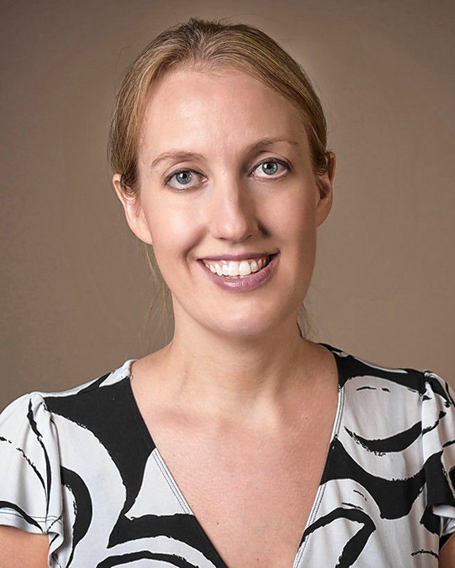
Elizabeth Clemente is a radiologist at Concord Imaging Center.


